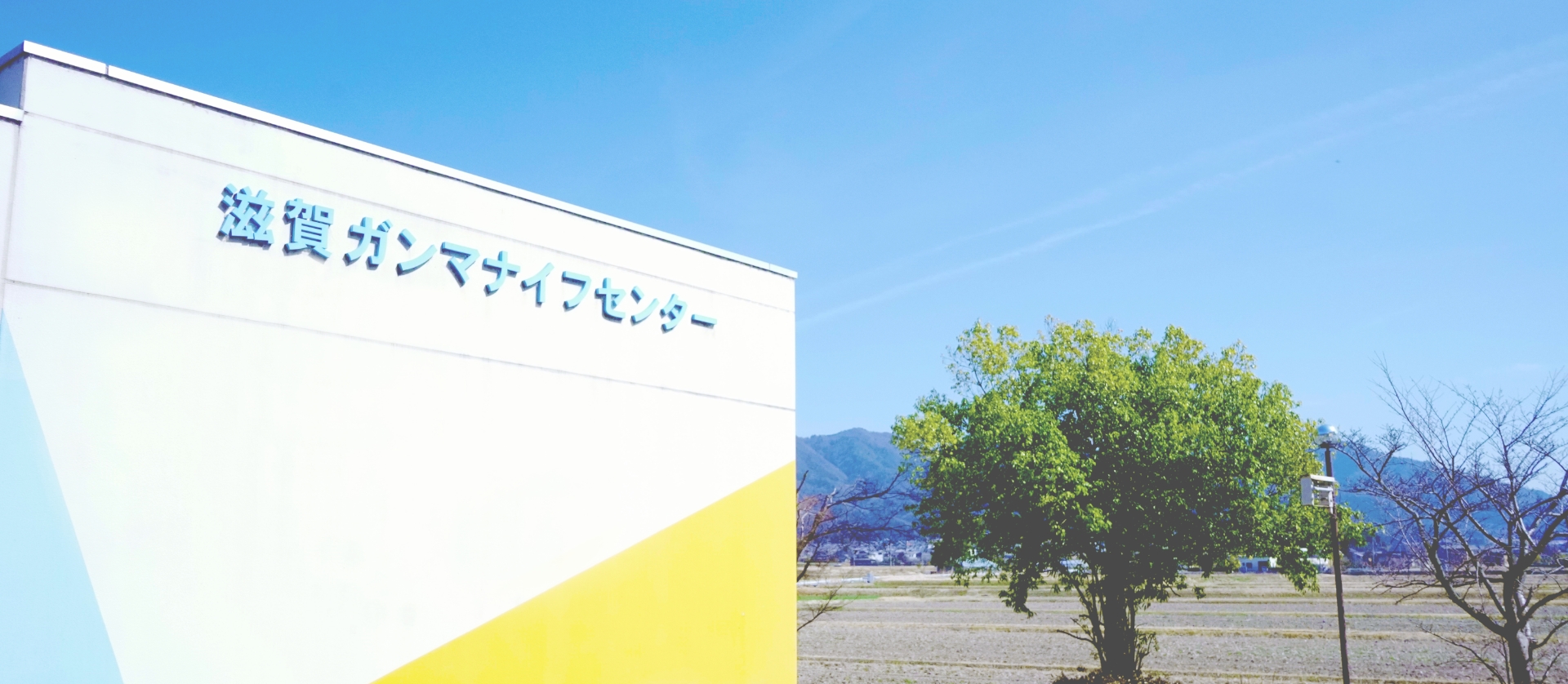
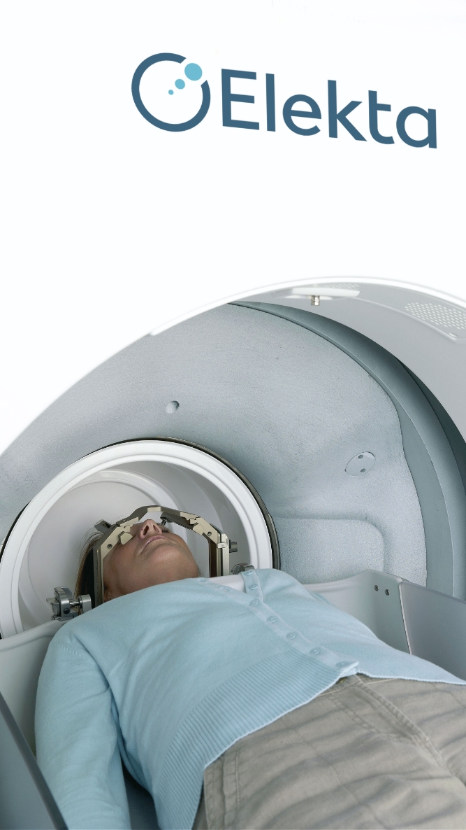
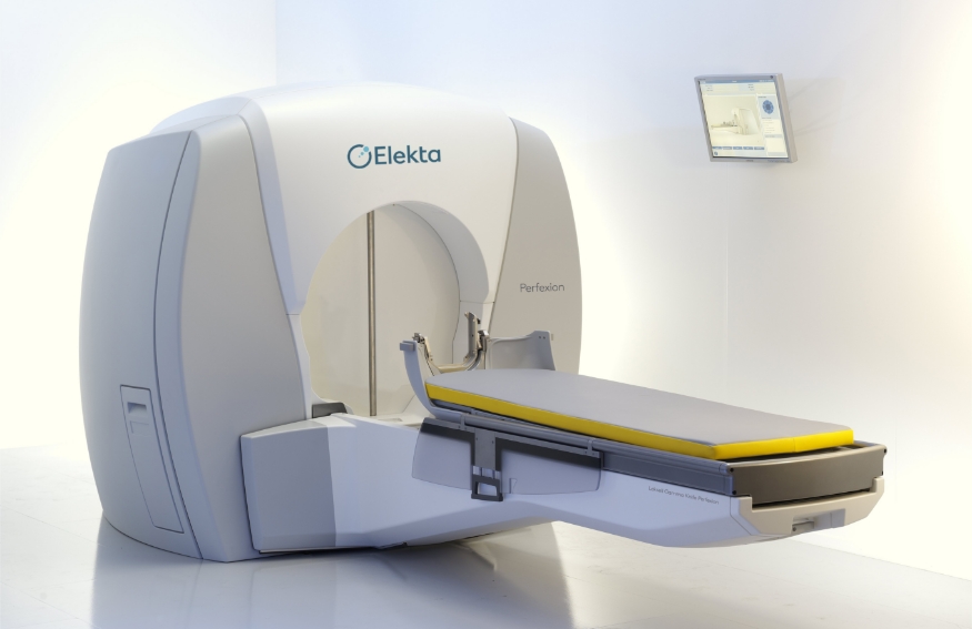
Shiga Gamma Knife Center is a specialized center located within Koto Memorial Hospital, offering Gamma Knife treatment, a form of stereotactic radio-surgery that is minimally invasive and can be performed in a relatively short time.
| Medical Facility | Koto Memorial Hospital |
|---|---|
| Address | 2-1 Hiramatsu-cho, Higashiomi, Shiga, Japan |
| Experience with Gamma Knife Treatment | Providing Gamma Knife treatment since 2004 |
| Hospital Website | www.koto-hp.jp |
| Gamma Knife Center Website | www.gammaknife-c.jp |
Gamma Knife treatment is provided by a team certified by the Japanese Society of Gamma Knife Radiosurgery. From online consultations before your trip to post-treatment follow-up, our physicians will guide you through each step of the process.
/ Specialist GK surgeon
A certified member of the Japan Leksell Gamma Knife Society team, providing Gamma Knife treatment.
She helps support international patients by guiding care coordination through treatment and follow-up.
/ Consultant Neurosurgeon
A certified member of the Japan Leksell Gamma Knife Society team, providing Gamma Knife treatment.
As part of a coordinated care team, he supports the process from pre-treatment review through post-treatment follow-up.
/ Consultant Neurosurgeon
With over 35 years of clinical experience, he has treated patients from more than 30 countries, including those requiring open cranial surgery. For Gamma Knife, he personally reviews your imaging (such as MRI scans) to confirm candidacy and clearly explain what to expect and the next steps.
Gamma Knife is a stereotactic radiosurgery (SRS) system that treats lesions in the brain by precisely focusing radiation on the target area. By delivering radiation from multiple directions and converging it on the lesion, it aims to control the lesion, including limiting further growth or reducing its size, while minimizing the impact on surrounding healthy brain tissue as much as possible.
Unlike open brain surgery, Gamma Knife treatment does not remove the lesion. Instead, it is considered a non-surgical treatment option, depending on factors such as the type, size, and location of the lesion.
In general, the process is as follows:
①placement of a dedicated head frame (Leksell frame),
②imaging studies (MRI and/or CT, and angiography if needed),
③treatment planning (GammaPlan),
④radiation delivery, and
⑤a follow-up examination the next day, with discharge in the morning if no problems are identified
For frame placement, local anesthesia is administered to the forehead and back of the head, and a dedicated metal frame is secured with four screws. If you feel particularly anxious, sedative medication may be used.
During treatment, patients generally do not feel pain or discomfort and can remain relaxed while lying down. The length of treatment depends on the size of the lesion and the radiation dose, but it typically takes around one hour on average. Bathroom breaks may also be available during the procedure if needed.