Conditions Commonly Treated
With Gamma Knife
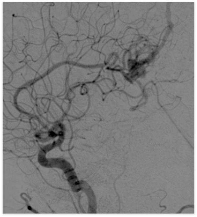
At our center, we provide Gamma Knife treatment primarily for brain tumors, and also for conditions such as arteriovenous malformations (AVMs) and trigeminal neuralgia. The first step is a physician review of your imaging studies (such as MRI) to determine whether Gamma Knife may be an appropriate option.
Vascular Conditions
- Arteriovenous malformation (AVM)
- Arteriovenous fistula (AVF)
- Cavernous malformation
(cavernous angioma), etc.
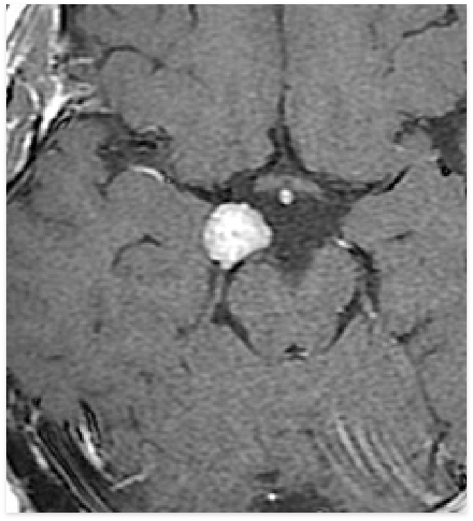
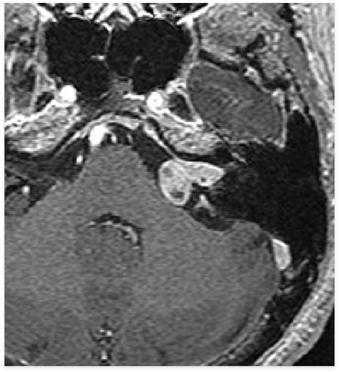
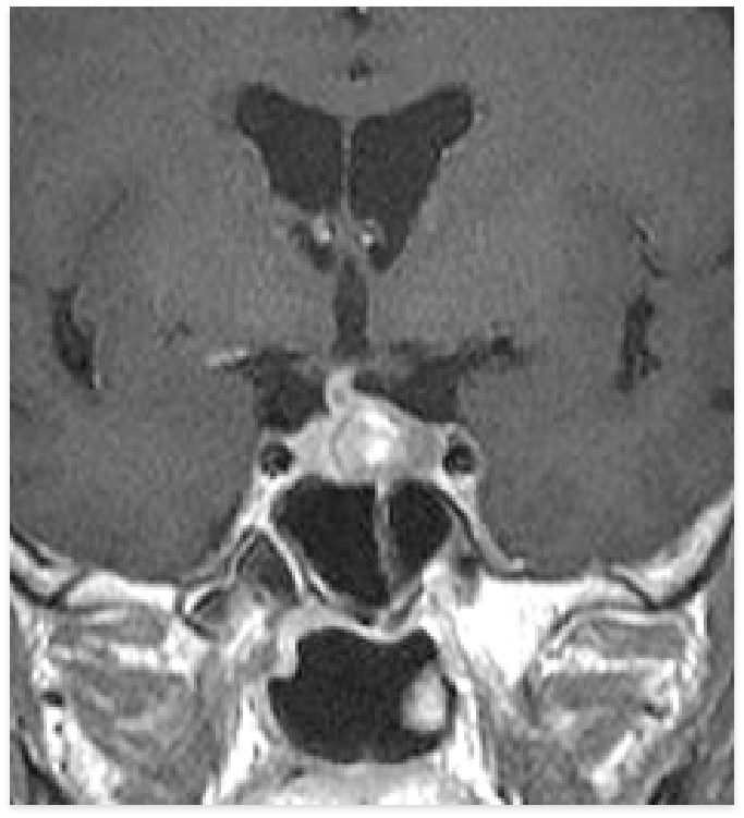
Benign Tumors
- Meningioma
- Vestibular schwannoma
(acoustic neuroma) - Pituitary adenoma
- Craniopharyngioma
- Chordoma
- Glioma, etc.
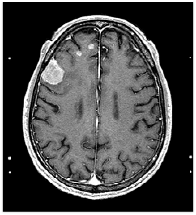
Malignant Tumors
- Brain metastases
(metastatic brain tumors) - Malignant glioma, etc.
Functional Disorders
- Trigeminal neuralgia
- Cancer-related pain
(not covered by insurance), etc.