2026.09.22
Contrast-Enhanced MRI: Why Is It Important for Brain Tumors and Gamma Knife?
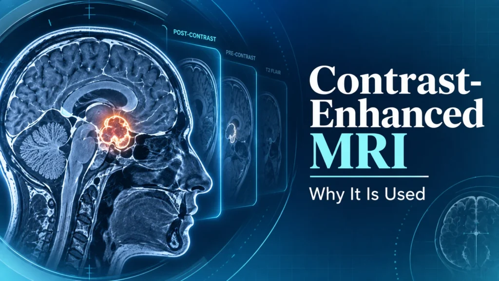
What Is Contrast-Enhanced MRI? What It Can Show in Brain Tumor Diagnosis
Contrast-enhanced MRI is an MRI scan in which a contrast agent is used to evaluate brain tumors, vascular lesions, and other conditions in more detail. For some lesions, using a contrast agent makes the borders and internal characteristics easier to see, providing important information for deciding on a treatment plan.
Why Contrast-Enhanced MRI Is Performed Before and After Gamma Knife Treatment
When considering whether Gamma Knife is appropriate, doctors look not only at the diagnosis but also at imaging to check the size, location, and number of lesions and their relationship to surrounding tissue. For brain metastases in particular, confirming the number and location of lesions in detail with contrast-enhanced MRI plays a role in treatment planning. MRI may also be used on the day of treatment to obtain images for planning.
However, not every patient necessarily needs the same MRI technique. The imaging methods and additional tests required may differ depending on the type of condition and treatment history. In addition, kidney function, a history of allergies, and other factors may call for an individual decision about whether to use a contrast agent. Please follow the instructions of your doctor or radiologist regarding whether contrast-enhanced MRI is needed.
Contrast-enhanced MRI is also used to evaluate the results after treatment. However, if a lesion looks larger on imaging, or the area of contrast enhancement changes, that alone does not necessarily mean the tumor has progressed. Post-treatment reactions, radiation necrosis, and other changes can produce similar-looking images, so comparison with previous images or additional tests may be needed.
If you are seeking a consultation about Gamma Knife from outside Japan, preparing the MRI images themselves, along with information showing the scan dates and the type of examination if possible, makes it easier for a specialist to assess the lesion. Imaging alone does not confirm whether you are a candidate for treatment; symptoms, treatment history, pathology results, and other factors are also evaluated.
Simply “having an MRI” is not necessarily enough for a pre-treatment evaluation for Gamma Knife. If the scans are old, or if they do not include the imaging needed to make treatment decisions, you may be asked to have an additional MRI. Especially when the lesion may be changing, new images may be needed to confirm its current status.
When sending images abroad, it is preferable to prepare, if possible, the original data saved as medical images rather than screenshots or photos. The required format, timing of scans, and type of examination differ by facility, so please check before sending. Imaging is important, but diagnosis and treatment candidacy cannot be determined from imaging alone.