2026.09.22
Follow-Up MRI After Gamma Knife: Why Is It Needed?
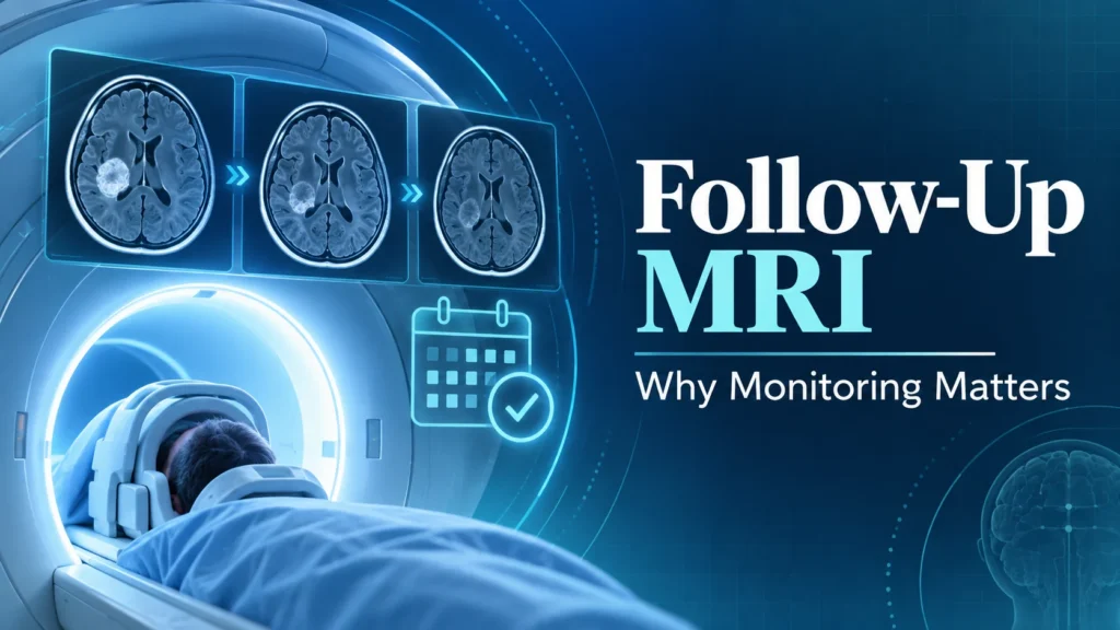
Why Is Follow-Up MRI Needed After Gamma Knife Treatment?
A follow-up MRI is an MRI scan performed to check for changes in the lesion and surrounding tissue after Gamma Knife treatment. Because Gamma Knife does not remove the lesion at the time of treatment, the effect of treatment is evaluated over time. Even if the lesion still appears on MRI after treatment, that alone does not mean the treatment is not working.
What Do Doctors Look For on MRI After Gamma Knife?
During follow-up, the new scan is compared with previous MRIs to see whether the treated lesion has shrunk, remained stable, or grown. At the same time, doctors also check for brain edema, treatment-related imaging changes, and the possibility of radiation necrosis. For brain metastases, it is also important to check not only the treated lesions but also whether new metastases have appeared elsewhere in the brain.
The timing and frequency of MRI scans vary depending on the type of condition, the lesion treated, symptoms, and how things have progressed so far. Gamma Knife Japan’s current guidance includes a recommendation for MRI follow-up every 6 to 12 months after treatment, but the same interval is not necessarily appropriate for every patient. Please follow your doctor’s instructions on when to have your scans.
For international patients, it is important to confirm before treatment where you will have MRI scans after returning home, in what format you will receive the image data, and who will evaluate the results. When you share images with the treatment facility to ask for its opinion, having both the previous and the latest images makes it easier to compare changes.
If new symptoms appear after treatment, you should not necessarily wait until your scheduled MRI. Depending on the nature and severity of the symptoms, an earlier medical visit or imaging test may be needed. It is important to plan your follow-up care to include not only “when to have an MRI” but also “where you will be seen” and “how your images will be shared.”
With follow-up MRI, it is especially important to be able to compare the new scan with previous images taken under the same conditions. If possible, keep the actual image data, not just the written report. Imaging methods can differ between countries and facilities, so if the treatment facility gives you instructions about how the scans should be taken, sharing them with your local medical facility makes comparison easier.
The course after Gamma Knife differs by condition. Benign tumors may require long-term monitoring, while for brain metastases, doctors check for new lesions in addition to evaluating the treated sites. For international patients, arranging before treatment to be able to continue having MRI scans after returning home is an important part of the treatment plan.